

The first line of cancer detection, which is accurate and early, is very important for the survival of a patient — and this is the point where the Dermoscopy Treatment figure shifts the entire diagnosis game. With providing a highly enhanced, clear, and lighted image of skin parts lying under the epidermis, dermoscopy is considered as the closest step to visual examination after biopsy.
It gives doctors the ability to find out the changes of cancer cells even before those changes can be seen by the naked eye, and thus they can give the patient a quicker, more exact, and less invasive type of treatment.
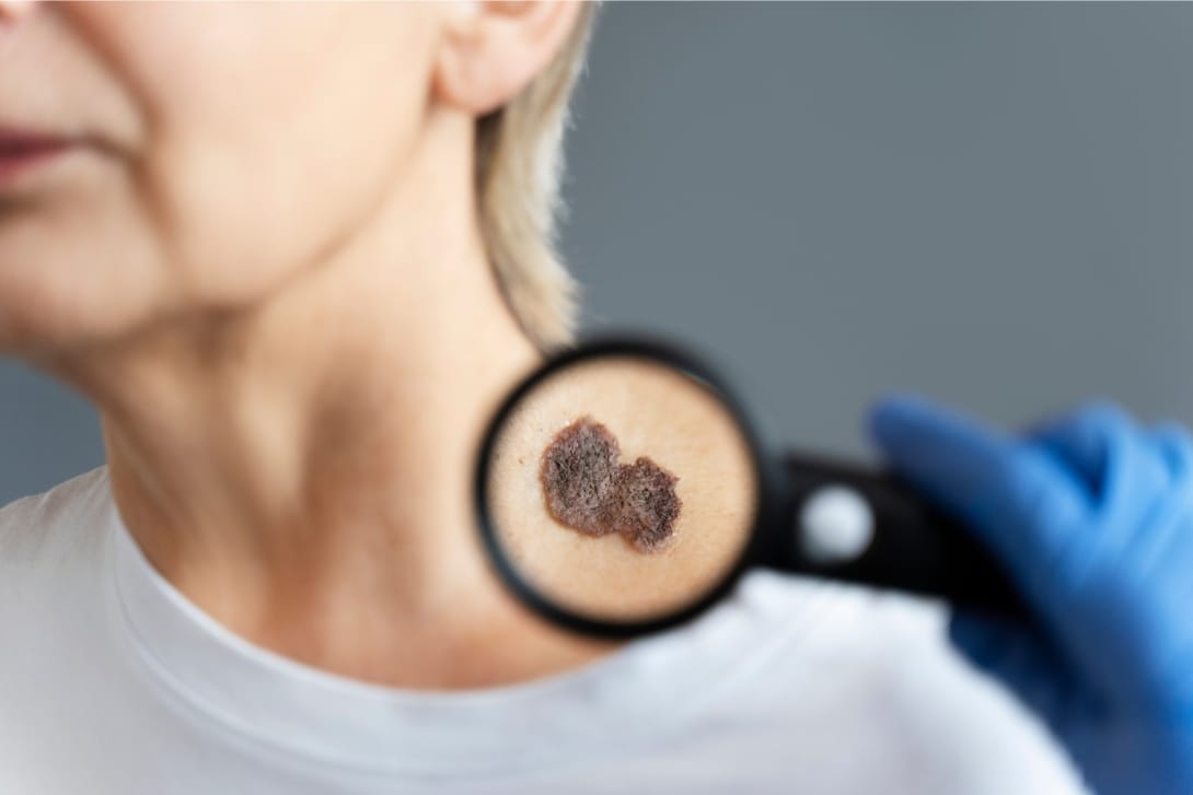
Understanding Dermoscopy: Seeing Beyond the Surface
Dermoscopy or dermatoscopy is a non-invasive diagnostic imaging technique that allows detailed examination of both pigmented and non-pigmented skin lesions. A dermatoscope is a device that uses magnification and polarized light to reveal skin patterns beneath the surface, which cannot be seen with normal light.
Dermatological research indicates that dermoscopy improves the diagnostic accuracy of melanoma by almost 30–40% as compared to the visual inspection of the skin alone. It can distinguish benign moles, abnormal lesions, and skin cancers in their earliest stages, thus, it is a must-have instrument in the dermatology of the present time.
How Dermoscopy Treatment Works
Dermoscopy Treatment refers to a procedure where a handheld dermatoscope or a digital imaging device is placed over the skin. The method is absolutely comfortable, secure, and lasts only a few minutes. Doctors look through the pigment network, blood vessel patterns, and structural asymmetries — these are the main features that show whether the lesion is cancerous or non-cancerous.
Basically, there are two different dermoscopy methods:
- Contact Dermoscopy: The instrument makes direct contact with the skin, usually with a liquid interface for better visualization.
- Non-Contact Dermoscopy: The use of polarized light removes the need for contact with the skin, thus guaranteeing a quick and clean examination.
Further innovations such as AI-powered dermoscopy have opened a way for dermatologists to use algorithms that can process hundreds of data points from the images to locate cancer at its earliest stage.
Dermoscopy for Skin Cancer: A Game Changer in Early Detection
Basically, the major benefit of a dermoscopy for skin cancer is the most proficient recognition of patterns. As a matter of fact, melanoma, basal cell carcinoma (BCC), and squamous cell carcinoma (SCC) are all characterized by different dermoscopic structures – just to name a few:
| Skin Cancer Type | Dermoscopic Clues |
| Melanoma | Asymmetric pigment network, irregular dots, blue-white veil |
| Basal Cell Carcinoma (BCC) | Arborizing vessels, translucent areas, shiny borders |
| Squamous Cell Carcinoma (SCC) | Scaly surface, blood spots, white structureless areas |
By identifying these early signs, dermoscopy for melanoma helps dermatologists detect cancer before it spreads deeper into the skin layers — where treatment becomes more complex.
Why Dermoscopy for Melanoma Matters
Melanoma is one of the most lethal types of skin cancer and causes about 75% of the deaths resulting from skin cancer. However, the survival rate after five years is as high as 98% when the cancer is detected at its early stage.
Using dermoscopy for melanoma, physicians can see the micro-patterns that are not visible to the human eye – very slight changes in pigment and blood vessel structures that point to cancer. Fewer unnecessary biopsies, faster patient reassurance, and timely treatment are the outcomes of this procedure.
Moreover, the use of digital dermoscopy instruments makes it possible to record changes in the skin over time which is an essential function for the follow-up of patients having a previous melanoma or atypical moles.
The Science Behind Dermoscopy Accuracy
Dermoscopy achieves its accuracy largely because it can go beyond the epidermal-dermal junction, which is the layer of the skin where a lot of melanocytic structures are found. With this help, the visualization can recognize pigment cell and blood vessel changes that cancer may have originated from.
In dermoscopy, the pictures taken can be computed by an algorithm that compares the shapes, symmetries, colors, and textures. The accuracy of AI-supported dermoscopy has been almost 95% for specific melanoma subtypes.
In addition, dermoscopic pictures being part of teledermatology can be sent confidentially for the opinion of an expert located remotely — thereby early detection getting facilitated even in those regions which lack proper medical facilities.
Dermoscopy vs. Traditional Examination
| Parameter | Traditional Visual Check | Dermoscopy |
| Precision | Limited to surface-level observation | Reveals subsurface structures |
| Invasiveness | May require biopsy for suspicion | Non-invasive, real-time insight |
| Time Taken | Dependent on lesion count | Quick, even for multiple lesions |
| Detection Accuracy | Moderate | Up to 40% higher accuracy |
Basically, Dermoscopy Treatment is a technique that provides clinicians with data from the patient’s body that confirms or denies their hypothesis before they do biopsies or excisions — thus, the number of these intervenes is lessened together with the reduction of the patient’s anxiety.
Integrating Dermoscopy into Preventive Skin Care
Consequently, the use of dermoscopy has become an integral part of preventive dermatology, which is done through yearly skin examinations, especially for people, who are most at risk, i.e. those with light skin, skin cancer in the family, or a history of getting a lot of sun.
Regular dermoscopy exams allow:
- First of all the detection of atypical lesions.
- The tracking of moles changing over time.
- Improved patient education and awareness.
- A visual archive for later comparison.
The trend is that dermatologists are more and more suggesting digital dermoscopy mapping which is a method that can find and keep the detailed photos of moles on the whole body and thus be able to follow even the smallest changes between the visits.
The Role of Technology: Digital & AI-Driven Dermoscopy
The present-day dermoscopy therapy is more than just a handheld tool. The integration with digital imaging, machine learning, and telemedicine is a way to improve diagnostic accuracy and patient experience.
- Incredible accuracy is achieved by AI algorithms when they are able to predict the probability of malignancy.
- 3D dermoscopy devices explore the lesions in detail, thus, revealing the structural irregularities that cannot be seen on 2D surfaces.
- A smartphone-compatible dermatoscopes allows remote consultations and early triage of patients who are living in distant areas.
Such innovations are redefining the role of dermoscopy as the hub of precision dermatology — a combination of clinical expertise and technological intelligence.
Safety, Comfort & Patient Benefits
Usually, patients are concerned about pain when diagnostic procedures are performed, however, dermoscopy is a treatment that is absolutely painless and without any risks. There is no radiation, incision, or recovery time. Actually:
- It is what causes the number of unnecessary biopsies to be reduced by 25–30%.
- The detection of melanoma is made up to 6 months earlier comparing to the visual exams.
- Increases patient confidence by providing them with a clear understanding of their lesions through visuals.
By this transparency, stronger doctor-patient relationships are being established which, in turn, lead to better long-term compliance with preventive skin care.
Real-World Impact: Statistics at a Glance
- By the time they are 70, one out of every five people is likely to get skin cancer.
- If the cancer is diagnosed early, the chances of survival go up from 65% to 98%.
- More than 80% of dermatologists worldwide rely on dermoscopy as their main screening method.
- The digital dermoscopy market is expanding at a CAGR of 11.2%, which is indicative of a rapid uptake in diagnostic care
In fact, these numbers strongly confirm the use of dermoscopy not only as a diagnostic instrument but also as a worldwide revolution for earlier, wiser, and safer cancer detection.
Skinstitute: Where Technology Meets Trust
At Skinstitute, we blend medical skill with the latest dermoscopic technology to provide precise, empathetic care. Our dermoscopy treatment services for skin cancer and melanoma are structured to locate alterations at the absolute earliest stage — thus, giving the patients the power of being clear and confident.
With the help of state-of-the-art digital dermoscopy devices, our doctors make the tailored diagnosis and after-care plans which combine medical science and the doctor’s empathy. At Skinstitute, we say every pixel counts because every life counts.
Key Takeaways
- Dermoscopy is a magnified, non-invasive technique that can be used to locate melanoma and skin cancer at an early stage.
- It basically upgrades the diagnostic accuracy by up to 40% and thus, helps in cutting down the biopsy tests that are needless.
- Dermoscopy of melanoma is very helpful in monitoring the changes of moles and recognizing the development of cancer at the earliest stages.
AI-assisted and digital dermoscopy instruments are leading a radical change in precision dermatology. - Such clinics as Skinstitute are not only redefining the concept of early skin cancer detection by combining advanced diagnostics with the provision of compassionate care but also setting new standards.
