- +91 7289828860
- contactwithskinstitute@gmail.com
- Nayapalli, Bhubaneswar-751015
- Home
- About Us
- Services
- Aesthetic Treatments in Bhubaneswar
- Botox Treatment
- Microbotox Treatment
- Hydrafacial Treatment
- Dark Circle Treatment
- Glutathione Treatment
- Thread Lift
- Vampire Lift
- Platelet Rich Plasma
- Chemical Peeling Treatment
- Micro Needling Treatment
- Low Level Laser Therapy With Face Mask
- Double Chin Reduction
- Dermal Fillers
- Exosomes Treatment
- Skin Booster Treatment
- Dermatosurgery Services in Bhubaneswar
- Clinical Dermatology
- Laser Skin Treatments in Bhubaneswar
- Trichology
- Venereology Treatment In Bhubaneswar
- Aesthetic Treatments in Bhubaneswar
- Gallery
- Blog
- Testimonials
- Home
- About Us
- Services
- Aesthetic Treatments in Bhubaneswar
- Botox Treatment
- Microbotox Treatment
- Hydrafacial Treatment
- Dark Circle Treatment
- Glutathione Treatment
- Thread Lift
- Vampire Lift
- Platelet Rich Plasma
- Chemical Peeling Treatment
- Micro Needling Treatment
- Low Level Laser Therapy With Face Mask
- Double Chin Reduction
- Dermal Fillers
- Exosomes Treatment
- Skin Booster Treatment
- Dermatosurgery Services in Bhubaneswar
- Clinical Dermatology
- Laser Skin Treatments in Bhubaneswar
- Trichology
- Venereology Treatment In Bhubaneswar
- Gallery
- Blog
- Testimonials
CONTACT US
- +91 7289828860
- Nayapalli, Bhubaneswar-751015
- contact@theskinstitute.in

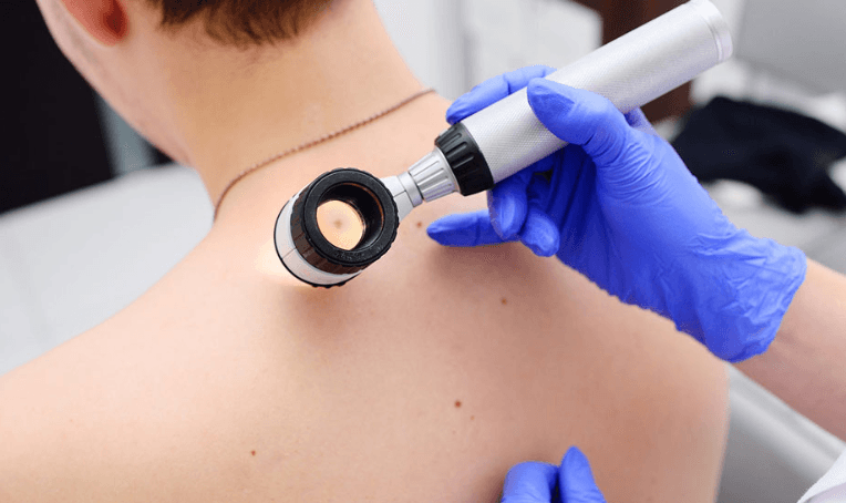
Are you worried about odd skin spots, recurring rashes, or moles? Our cutting-edge dermoscopy services at The Skinstitute in Bhubaneswar provide a non-invasive and incredibly precise way to evaluate skin abnormalities. Our skilled dermatologists can see beneath the surface of the skin in real time by using a dermatoscope, a portable magnifying tool with polarized light. This aids in the early identification of vascular lesions, fungal infections, inflammatory diseases, and skin cancers (such as melanoma). In many situations, it supports accurate, early-stage diagnosis and does away with the need for immediate skin biopsies. The process provides clarity and peace of mind in a timely and efficient manner and is safe, painless, and usually finished in 20 to 30 minutes.
Treatment Overview
By enabling a closer look beneath the skin’s surface, dermoscopy—also referred to as dermatoscopy or epiluminescence microscopy—improves diagnostic precision. Dermoscopy types include:
The best way to see pigment networks, vascular patterns, and deeper skin structures is with polarized dermoscopy.
The best method for examining surface characteristics like crusts, scaling, and superficial pigmentation is non-polarized dermoscopy.
Digital dermoscopy is the process of taking and saving high-resolution pictures in order to track skin lesions over time.
Contact dermoscopy: To improve clarity and lessen skin reflection, direct contact and a liquid medium are used.
Why Choose The Skinstitute for Dermoscopy Treatment & Its Key Benefits?
- Assessment under the supervision of a doctor by licensed dermatologists with extensive dermoscopy training.
- Early identification of skin cancers, including basal cell carcinoma and melanoma.
- Helps prevent needless skin biopsies.
- Uses digital dermoscopy to track changes in moles over time.
- Useful in the diagnosis of inflammatory dermatoses, pigment disorders, and infections.
- Safe for all skin types and tones.
- In-clinic process that is quick and painless.
Who is it For? & How It Works (Ideal Candidates)
Dermoscopy is best for people with:
- Changing or irregularly shaped moles
- Have persistent or inexplicable rashes
- Observe any vascular lesions or skin discolorations.
- They are susceptible to skin cancer due to sun exposure and family history.
- Long-term skin growth monitoring is required.
The dermatologist can observe skin patterns and colors that are invisible to the human eye by illuminating the skin and magnifying it up to 10–20 times. This facilitates prompt, accurate diagnosis without the need for surgery.
Step-by-Step Procedure: Dermoscopy Treatment At The Skinstitute
Before Your Visit
- Consultation & Skin Assessment: The necessity of a dermoscopy will be discussed, and your skin concern will be assessed.
- Instructions for Pre-care: Just clean, makeup-free skin is required.
During Procedure
- Procedure & Timeline: The lesion site is examined using a dermatoscope, either with or without contact fluid. Usually, the process takes 20 to 30 minutes.
- Comfort Management: Painless and non-invasive. No numbing or anesthesia is necessary.
Aftercare & Safety Downtime
- Advice for After the Procedure: Avoid downtime. Get back to your regular activities right away.
- Results Timeline: If additional assessment or treatment is necessary, immediate insights are frequently available.
Dermatologists utilize dermoscopy, sometimes referred to as dermatoscopy or epiluminescence microscopy, as a non-invasive diagnostic technique to look more closely at skin abnormalities. It makes use of a portable instrument known as a dermatoscope, which lights and magnifies skin lesions to reveal details about their internal architecture that are invisible to the unaided eye. When it comes to the diagnosis and treatment of different skin disorders, dermoscopy has several advantages.
- Enhanced Visualization: Dermatologists can see things like pigment patterns, vascular patterns, and other features inside skin lesions that are invisible to the unaided eye thanks to dermoscopy, which magnifies the image of the lesions. Accurate diagnosis of skin disorders, such as melanoma, basal cell carcinoma, and other forms of skin cancer, is made easier by this improved visibility.
- Improved Diagnostic Accuracy: By enabling dermatologists to assess the morphological features of skin lesions in detail, dermoscopy helps improve diagnostic accuracy and distinguish between benign and malignant lesions. Dermoscopic criteria for different skin conditions have been established, allowing for more precise diagnosis and appropriate management.
- Monitoring of Skin Lesions: Dermoscopy is a useful tool for tracking the evolution of skin lesions over time. Dermatologists can monitor the development of lesions, evaluate the effectiveness of treatment, and identify early indicators of disease recurrence by recording dermoscopic features and comparing images during follow-up examinations.
- Facilitation of Dermatologic Procedures: Dermoscopy can assist dermatologists in performing various dermatologic procedures, such as skin biopsies and surgical excisions. By guiding the selection of biopsy sites and delineating lesion borders, dermoscopy helps ensure accurate sampling and precise removal of suspicious lesions, minimizing the risk of incomplete excision or unnecessary procedures.
- Patient Education and Counseling: Dermoscopy allows dermatologists to visually communicate diagnostic findings and treatment recommendations to patients, enhancing patient understanding and involvement in their care. Visual documentation of skin lesions using dermoscopy images can also serve as a useful educational tool for healthcare providers and patients alike.
Overall, dermoscopy is a useful additional tool for dermatologists, providing better visibility, more accurate diagnosis, early skin cancer detection, and better skin lesion monitoring. The assessment and treatment of numerous dermatological disorders have been transformed by its incorporation into clinical practice, eventually improving patient outcomes and care.

Before

After
What People Says
EXCELLENTTrustindex verifies that the original source of the review is Google. Awesome treatments.... Worth it..🥳🥳Posted onTrustindex verifies that the original source of the review is Google. Abhishek Sir was truely a blessing in terms of saving my hair and helped a lot for growing my hair again and also helped regarding my acne issuePosted onTrustindex verifies that the original source of the review is Google. Best Dermatologist.Posted onTrustindex verifies that the original source of the review is Google. Visiting SKINSTITUTE and consulting Dr Kumar Abhishek is absolutely praise worthy as the Dr's diagnosis and advises are excellent. I and family members are getting excellent result by visiting such a nice doctor.Posted onTrustindex verifies that the original source of the review is Google. Better place to cure your skin related issues. Satisfactory and cure level-100% Most recommended ❤️❤️❤️Posted onTrustindex verifies that the original source of the review is Google. Good experience, treatment is very goodPosted onTrustindex verifies that the original source of the review is Google. I was struggling with my psoriasis for a long time,but after meeting Dr Kumar Abhishek, Finally started to change for the better.His clear understanding of the condition, patient approach,and effective treatment plan gave me both relief and hope.I am truly grateful for his guidance and carePosted onTrustindex verifies that the original source of the review is Google. Nice doctor...... Staff behavior so nice... Very satisfied indeedPosted onTrustindex verifies that the original source of the review is Google. The doctor and staff have very well manner and helpful. Also, I have seen the improvement in my skin as well.
Frequently Asked Questions (FAQ)
Dermatologists can identify asymmetries and irregular features that are frequently observed in melanoma in its early stages by using dermoscopy, which enables visual evaluation of pigment patterns and vascular structures.
Yes, dermoscopy works well and is safe for all skin types. It assists in identifying minute alterations and pigment changes that might go unnoticed during a routine skin examination.
Of course. In order to distinguish infections from dermatitis or psoriasis, dermoscopy provides information on pigmentation, vessel configurations, and scaling patterns.
Depending on how many lesions are inspected, a dermoscopy session typically lasts between 20 and 30 minutes.
Yes, in a lot of instances. It can reduce the need for skin biopsies by non-invasively ruling out malignancies. Nonetheless, a biopsy might still be recommended in questionable situations.
Annual examinations are recommended for people with multiple or atypical moles, or more frequently if skin cancer runs in the family.
Indeed. It is frequently used to check children for skin infections or birthmarks and is totally safe for pediatric cases.
Indeed. The diagnosis of rosacea and its subtypes is supported by vascular patterns such as telangiectasia that can be seen during dermoscopy.
Reporting changes is necessary. Digital dermoscopy makes it possible to compare results over time and identify even slight changes.
If the doctor thinks it’s necessary, it’s part of the dermatological evaluation in most clinics, including The Skinstitute.


